Ever wondered how doctors can see what’s going on inside your body without making a single cut? The answer is diagnostic imaging. In simple terms, these are the techniques used to create detailed pictures of your internal organs, bones, and tissues. Think of it as a set of highly advanced tools that let medical professionals play detective, looking for clues inside you.
A Clearer Look Inside Your Body
So, what exactly is diagnostic imaging? It's a field of medicine that uses different forms of energy—like X-rays, sound waves, or magnetic fields—to produce incredibly detailed images of your anatomy. These pictures are fundamental to modern healthcare, giving doctors a window into what’s causing your symptoms.
Imagine trying to fix a complex engine without being able to look under the bonnet. You could guess what’s wrong, but you’d never be certain. Diagnostic imaging lifts that bonnet, giving clinicians a direct view to identify problems with remarkable precision. This visual information is crucial for making an accurate diagnosis, monitoring a condition over time, or planning a medical procedure.
The Role of Imaging in Modern Medicine
The importance of these scans in day-to-day healthcare can't be overstated. In the UK, the NHS relies heavily on imaging to manage patient care effectively. To give you an idea of the scale, in the 2024/25 fiscal year, NHS England reported performing over 41 million diagnostic imaging tests. That number alone shows just how central these procedures are.
This field is also moving forward at a rapid pace. New methods are making scans faster, safer, and more accurate than ever before. You can explore the latest breakthroughs in our guide to advancements in diagnostic technologies.
At its core, diagnostic imaging replaces guesswork with evidence. It empowers doctors with the visual proof needed to diagnose illnesses earlier, track the effectiveness of treatments, and guide interventions like surgery with pinpoint accuracy.
An Overview of Common Scans
To give you a better idea of the tools in the toolbox, here’s a quick guide to the most common imaging types. Each one works differently, making it suited for answering specific medical questions.
Quick Guide to Common Diagnostic Imaging Types
This table breaks down the main types of diagnostic imaging, how they work, and what they're most commonly used for. It’s a handy reference to see which scan is right for which job.
Each of these scans offers a unique perspective, allowing doctors to choose the best possible method to get the answers they need for your care.
Exploring the Main Types of Medical Scans
Ever wondered why a doctor might order an MRI for a knee injury but an X-ray for a suspected broken arm? The choice isn't random. It’s a precise decision based on what each type of diagnostic imaging does best, with different scans using different technologies to see inside the body, each offering a unique perspective.
Understanding these differences helps to demystify the process and makes you a more informed patient. Let’s break down the most common types of medical scans, exploring how they work and where they fit in real-world diagnosis.
This map shows how imaging isn't just about finding problems. It also plays a crucial role in monitoring health over time and guiding treatments to be as precise and effective as possible.
X-rays: The Foundation of Medical Imaging
The X-ray is often the first scan people think of, and for good reason. It’s one of the oldest and most frequently used forms of medical imaging.
An X-ray machine sends a small, controlled dose of ionising radiation through the body. Dense materials like bone absorb more of this radiation, so they appear white on the final image. Softer tissues, like muscle and fat, let more of the radiation pass through, showing up in shades of grey.
This simple but effective principle makes X-rays brilliant for:
- Detecting Fractures: They provide sharp, clear images of bones, making it easy to spot breaks or dislocations.
- Examining the Chest: A chest X-ray can reveal lung conditions like pneumonia or a collapsed lung.
- Screening Mammograms: Specialised X-rays are used to check for signs of breast cancer.
CT Scans: A 3D View of the Body
A Computed Tomography (CT) scan takes the concept of the X-ray a big step further. Instead of a single flat image, a CT scanner takes a series of X-ray images from many different angles around the body. A computer then processes these images to create detailed, cross-sectional "slices."
Think of it like looking at a single slice of a loaf of bread versus seeing the whole loaf from the outside. The slice gives you a much better view of the internal texture. These slices can even be combined to create 3D models, giving doctors an exceptionally detailed look at bones, organs, and blood vessels.
CT scans are the go-to for complex situations. They are invaluable in emergency medicine for quickly assessing internal injuries from trauma and are also used to diagnose tumours, blood clots, and internal bleeding.
MRI Scans: Detailed Soft Tissue Imaging
Unlike X-rays and CT scans, a Magnetic Resonance Imaging (MRI) scan uses no ionising radiation. Instead, it relies on a powerful magnetic field, radio waves, and a computer to generate incredibly detailed pictures of organs and soft tissues.
The machine’s strong magnet aligns the protons in your body's water molecules. Radio waves are then used to briefly knock these protons out of alignment. As they snap back into place, they emit signals that the MRI scanner detects and uses to build an image.
Because different tissues realign at different rates, an MRI can distinguish between them with remarkable clarity. This makes it the top choice for:
- Joint and Ligament Injuries: An MRI can clearly show tears in tendons, ligaments, and cartilage.
- Brain and Spinal Cord Issues: It's excellent for detecting tumours, strokes, and conditions like multiple sclerosis.
- Organ Examination: It provides detailed views of organs like the liver, heart, and kidneys.
The demand for these advanced scans has risen sharply. In England, between 2016/17 and 2023/24, CT scan activity increased by about 35%, and MRI scans grew by nearly 40%. This shows just how important they've become in diagnosing complex health conditions.
Ultrasound Scans: Real-Time Imaging with Sound
Ultrasound imaging, also known as sonography, uses high-frequency sound waves to create live images from inside the body. It’s the same basic technology used in naval sonar, just adapted for medical use. A small handheld device called a transducer emits the sound waves and detects the echoes that bounce back from tissues and organs.
A computer translates these echoes into a real-time image on a screen. Because it uses no radiation, it's considered extremely safe and is the standard for monitoring pregnancies.
But ultrasound is incredibly versatile. It's used for everything from looking at gallstones to guiding injections. One of the best examples is the heart echocardiogram, and this a friendly guide to heart echocardiograms explains how it works. This specific type of ultrasound creates moving pictures of your heart, assessing its chambers and valves. To learn more, you can read our own guide on what to expect during a heart echo scan.
How to Prepare for Your Imaging Appointment
Knowing what to do before your diagnostic imaging appointment can make the whole experience feel smoother and less stressful. While preparation is usually straightforward, the specifics can change quite a bit depending on the type of scan you’re having.
We'll walk you through the essential steps, from general advice that applies to every scan to the detailed instructions you'll need for specific procedures.

Being prepared empowers you and helps our clinical team get the clearest possible images. A little planning really does go a long way.
Universal Preparation Tips for Any Scan
No matter which scan you're scheduled for, a few universal tips will help your appointment go off without a hitch.
First, always bring your referral letter and any other relevant medical documents. It’s also a good idea to have a list of your current medications and any known allergies. This information is vital for the clinical team to ensure your safety.
Before you arrive, it's smart to have a few questions ready for your doctor or the imaging centre. Never hesitate to ask about anything you're not sure of.
A few key questions to ask beforehand:
- How long will the entire appointment take?
- Are there any specific dietary rules I need to follow?
- Will I be able to drive myself home afterwards?
- When and how should I expect to receive my results?
Getting answers to these questions helps you plan your day and can ease any anxiety you might be feeling.
Scan-Specific Instructions You Need to Know
While the general tips are a great starting point, different imaging techniques require their own unique preparations. Following these instructions carefully is essential for the quality of the scan and your safety.
For an MRI scan, the single most important rule is to remove all metal from your body. The scanner’s powerful magnet can turn metallic objects into dangerous projectiles. This means jewellery, watches, hearing aids, and even clothing with metal zips or underwires. You must also tell the radiographer about any internal metal, like a pacemaker, cochlear implant, or surgical clips.
An ultrasound scan of your abdomen or pelvis often requires some simple but crucial preparation. For an abdominal scan, you might be asked to fast for several hours to reduce gas in your intestines, which can obscure the view. For a pelvic ultrasound, you'll likely need to drink several glasses of water and hold it in, as a full bladder provides a clear "window" for the sound waves to pass through.
For some CT scans, particularly those looking at the abdomen, you may need to fast for a few hours. This ensures the images are as clear as possible, without any interference from food in your digestive system.
Remember: These specific instructions aren't just suggestions; they are crucial for obtaining accurate diagnostic images. If you are unsure about any instruction, always call the imaging centre in advance to clarify.
Understanding Contrast Agents
For certain MRI or CT scans, your doctor might recommend using a contrast agent. This is a special substance, usually given via an injection, that helps to highlight specific organs, blood vessels, or tissues. It makes them show up more clearly and in greater detail on the final images.
Think of it as a temporary dye that makes certain areas of your body "glow" for the scanner.
These agents are generally very safe and are naturally flushed out of your body within a day or so. When the contrast is injected, you might feel a brief warm or flushing sensation, or maybe a metallic taste in your mouth. This is completely normal and passes quickly.
To get a better sense of the process, you can learn more about what to expect from MRI scans with contrast in our detailed guide. Understanding what these agents are and why they're used can make the experience much less daunting.
What to Expect During and After Your Scan
Once you’ve done all the prep, you’re ready for the scan itself. Knowing what the experience will actually feel like can go a long way in calming any last-minute nerves. It’s also helpful to understand what happens after you walk out of the imaging suite, so you’re clear on the next steps. The journey isn’t really over until you and your doctor have the answers you came for.
Let's walk through what you can expect, from the moment you step into the scanning room to the day you get your results.
Inside the Scanning Room
The environment and what you experience will vary quite a bit depending on the type of scan you're having. A radiographer will be with you every step of the way, making sure you’re comfortable and positioned just right to get the clearest possible images.
For an MRI scan, you’ll lie on a motorised bed that slides into a large, tunnel-like machine. The first thing most people notice about an MRI is the noise—it makes a series of loud, repetitive knocking and buzzing sounds. We’ll give you earplugs or headphones to block it out. The most important thing is to lie as still as you can to avoid blurring the images.
A CT scan also uses a motorised bed, but it moves through a large, ring-shaped machine that looks a bit like a giant doughnut. It's a much faster and quieter process than an MRI. You might feel the bed move in small increments as the scanner works, but the procedure itself is completely painless. To get a better idea of timings, you can read our detailed guide on how long a CT scan takes.
No matter which scan you’re having, the radiographer is in constant contact with you through an intercom. You're never alone, and they are right there to support you if you have any questions or concerns during the procedure.
Comparing Patient Experiences MRI vs CT Scan
To give you a clearer picture, here’s a quick comparison of what patients typically experience during these two common scans. It really helps to know the key differences in how long they take, what they sound like, and how they feel.
Understanding these points beforehand can make the entire process feel much more manageable and less intimidating.
After the Scan: The Path to Your Results
Once the scan is finished, your job is pretty much done. Unless you've had a sedative, you can usually get back to your day right away. Now, the behind-the-scenes work kicks in.
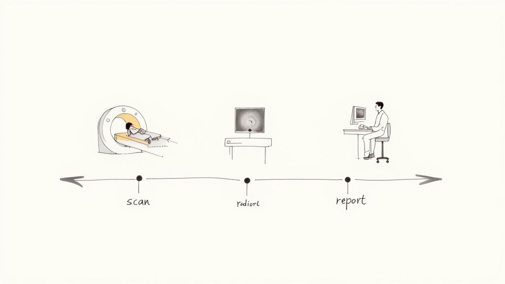
The images from your scan aren't just simple pictures; they're incredibly detailed sets of data that require a specialist's eye. This is where a radiologist comes in. A radiologist is a doctor who has spent years training to interpret medical images to diagnose and monitor injuries and diseases.
They will meticulously review your scans, looking for any abnormalities or signs of a condition. The radiologist then compiles their findings into a detailed report, which is sent directly to the doctor who referred you in the first place.
Receiving and Discussing Your Findings
This final step is probably the one you’ve been waiting for. Your referring doctor, who knows your full medical history, will receive the radiologist's report, usually within a few business days.
Your doctor will then schedule a follow-up appointment to go over the results with you. This is your chance to really understand what the images revealed, ask any questions you have, and talk about what comes next for your care plan.
Waiting for any kind of medical result can be tough. If you're curious about typical timelines for diagnostic information, you might find some useful context in resources about understanding how long to expect results for other common medical tests. It can help set realistic expectations for the whole process.
Navigating Safety and Risks in Medical Imaging
It’s completely natural to have questions about the safety of any medical procedure, and diagnostic imaging is no different. Understanding the risks—however small—is a key part of feeling confident in your care.
The good news is that medical imaging is a highly regulated and overwhelmingly safe field. The simple truth is that the diagnostic benefits almost always outweigh the risks.
Every scan is performed with a principle known as ALARA in mind, which stands for "As Low As Reasonably Achievable." This golden rule ensures that clinicians use the absolute minimum dose of radiation needed to capture high-quality images for a precise diagnosis. Your safety is always the first priority.
Understanding Radiation in Context
When people hear the word "radiation," it can sound a bit alarming. But it’s important to remember that we’re all exposed to a small, safe amount of natural radiation every single day from our environment. This is what’s known as background radiation.
Putting the radiation from an X-ray or CT scan into this context is really helpful. The dose from a single chest X-ray, for example, is often compared to the amount of natural background radiation you’d get over just a few days of normal life.
A more powerful CT scan might deliver a radiation dose equivalent to a few years of background radiation. While that's higher, it's still considered a very low risk, especially when weighed against the critical information the scan can provide to diagnose a serious condition.
This managed approach ensures that the medical benefit is always the clear priority.
The Safety of Different Scan Types
Each type of diagnostic imaging has its own specific safety profile, which is why your doctor chooses a particular scan for your situation. It's all about balancing the need for information with your wellbeing.
Here's a quick breakdown of what to expect from the main types of scans:
- X-rays and CT Scans: These use ionising radiation, but the doses are carefully controlled. The risk is extremely low, and these scans provide invaluable information, particularly for bone injuries and complex internal conditions.
- MRI Scans: An MRI uses a powerful magnet and radio waves, not ionising radiation. Here, the primary safety focus is on the magnet. Strict rules prevent any metal objects from entering the scan room, and patients with certain metallic implants may not be able to have an MRI.
- Ultrasound Scans: Ultrasound is considered one of the safest imaging methods out there. It uses sound waves, not radiation, which is why it's the standard choice for monitoring pregnancies without any known risk to the mother or baby.
Your Safety Is the Top Priority
Ultimately, every diagnostic imaging procedure is performed under the close supervision of highly trained professionals, including radiographers and radiologists. They are experts in keeping you safe while capturing the clearest possible images.
Before any scan involving radiation, the clinical team carefully weighs the potential benefits against the tiny potential risks. This means that when a scan is recommended, it’s because your doctor has decided it’s the best way forward to get the answers needed to manage your health effectively. You are always in safe hands.
Accessing Diagnostic Imaging Services in the UK
When you need a medical scan in the UK, the first step is figuring out which route is right for you. Your path will usually depend on how urgent your situation is and what you’re most comfortable with. Essentially, there are two main ways to get the scan you need: through the National Health Service (NHS) or via private healthcare.
No matter which option you choose, the journey almost always starts with your GP or a specialist. If they believe a scan is medically necessary, they'll give you a referral. Think of this referral as your key—it unlocks access to imaging services, whether you stick with the NHS or decide to go private.
The NHS Pathway
Once you have an NHS referral, you’ll be put on a waiting list for your scan at a local hospital or one of the new community diagnostic centres. The quality of care is fantastic, but the waiting times can be a real sticking point. They vary hugely depending on the type of scan you need and how much demand there is in your area. This is the standard path for most people, and it’s all covered by the NHS.
The Private Healthcare Alternative
For anyone looking for a faster answer, the private route is a very compelling alternative. You can take the exact same referral from your GP and use it to book an appointment at a private hospital or a specialist diagnostic centre like The Vesey. The big difference here is speed—you can often get a scan scheduled within days, not weeks or months.
Opting for a private centre comes with some clear benefits:
- Speed: Waiting times for appointments and results are drastically shorter.
- Advanced Technology: You often get access to the very latest imaging equipment, which means clearer, more detailed scans.
- Specialised Staff: Your scan will be handled by highly experienced radiographers and radiologists.
- Patient Experience: The whole process tends to feel more streamlined, comfortable, and personal.
The private diagnostic sector is a significant and growing part of the UK's healthcare landscape. Opting for a private scan means bypassing NHS waiting lists and gaining direct access to specialised care when you need it most.
Making an Informed Decision
The choice between the NHS and private healthcare is a personal one. The NHS delivers essential, high-quality care to millions. Where the private sector really shines is in its speed, convenience, and access to the newest technologies. This thriving market, valued at around £2.3 billion in 2024, is all about giving patients more control over their healthcare journey. This growth also fuels healthy competition, pushing for constant improvements in service and technology across the board. You can discover more insights about the UK's diagnostic imaging market on ibisworld.com.
Of course, cost is a major factor for most people. To give you a clearer idea of the investment involved, you might find it helpful to read our guide on how much a private MRI scan costs. At the end of the day, whether you go through the NHS or a private clinic, the goal is exactly the same: to give you and your doctor the clear, accurate information needed to make the best decisions for your health.
A Few Common Questions About Medical Scans
Even after getting to grips with the different types of diagnostic imaging, it's completely normal to have a few more questions rattling around. We get it. Here are some quick, clear answers to the queries we hear most often, designed to give you that extra bit of clarity and peace of mind.
Are Medical Scans Painful?
The short answer is no, not really. For the most part, diagnostic imaging is completely painless. Scans like X-rays, CTs, and ultrasounds are non-invasive, which just means nothing actually enters your body. You might be asked to hold a slightly awkward position for a moment, but you won't feel the scan itself.
An MRI is also painless. Some people find lying still in the scanner a bit strange or claustrophobic, and it can be quite noisy—which is why we always provide ear protection. If your scan requires a contrast agent, you might feel a tiny, brief pinprick from the injection, but it’s over in a second.
The most common feedback we get is that the thought of the scan is often far worse than the experience itself. Our team is right there with you, making sure you're as comfortable as possible from start to finish.
How Long Until I Get My Results?
The wait for results can be a nerve-wracking time, so it helps to understand what’s happening behind the scenes. Once your scan is done, the images are sent to a radiologist—a specialist doctor—who meticulously analyses every detail. They then compile their findings into a detailed report for your referring doctor.
This whole process usually takes a few business days. Your own doctor, who knows your full medical history, will then get in touch to arrange a follow-up appointment. They’ll walk you through the results and explain what the next steps are for your care.
Can I Have a Scan If I’m Pregnant?
Your safety, and that of your baby, is always our absolute priority. Scans that don’t use ionising radiation, like ultrasound and MRI, are generally considered safe during pregnancy. In fact, ultrasound is the main tool we use to check on a baby's development.
We typically avoid scans that involve radiation, such as X-rays and CT scans, unless there's a significant medical reason where the benefit clearly outweighs any potential risk. Your doctor will always have a detailed conversation with you about this, ensuring you make the safest possible choice together.
What Exactly Does a Radiologist Do?
Think of a radiologist as a highly trained medical detective. They are specialist doctors who have completed years of extra training to become experts at interpreting medical images.
They don’t just glance at a picture; they dive deep into the complex data from your scan to spot abnormalities, diagnose conditions, and monitor how a treatment is working. Their expert report gives your doctor the critical information needed to make the right decisions about your health.
At The Vesey, we believe in clear communication and getting you the answers you need, quickly. Whether you have a GP referral or want to book a private consultation, our team is here to guide you every step of the way. To find out more or to book an appointment, please visit us at https://www.thevesey.co.uk.
CQC-regulated private hospital in Sutton Coldfield. 25+ specialties, 68 expert clinicians. Open 7 days, 8am–8pm. No waiting lists. Free parking.
Book an appointment